Video Article Open Access
Is Micromachined Back-Markers on Thin Film of Scaffold Effective to Measure Repetitive Contraction of Myotubes?
Shigehiro Hashimoto
Kogakuin University, Tokyo, 1638677, Japan
Vid. Proc. Adv. Mater., Volume 2, Article ID 2108214 (2021)
DOI: 10.5185/vpoam.2021.08214
Publication Date (Web): 25 May 2022
Copyright © IAAM
Graphical Abstract
Abstract
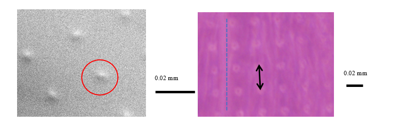
Most of biological cells adsorb on the scaffold, and show activities: migration, deformation, proliferation, and differentiation. These activities depend on properties of the scaffold. The micro topography of the surface of the scaffold, which is close to the cell size, is effective for several applications [2]: the marker to trace each cell, and the tool to control the activity of each cell. The cell aligns along the micro step lower than 1 μm [3]. The micro-striped groove can control the cell orientation in the flow channel. The aspect ratio of the checkered concexo-concave pattern can control the orientation of cells. The taper-striped pattern is effective to observe durotactic migration of cell [4]. C2C12 (mouse myoblast) is used in the present study. The typical diameter of the cell is 20 μm, when it is floating in the medium. The scaffold of the transparent film with micro pattern markers has been designed to measure the contractile force of myotube under electric stimulation in vitro. The scaffold is made of a polydimethylsiloxane thin film (thickness 6 μm), of which the back side has arrangement of micro-protrusions (4 μm diameter, 2 μm height, interval 30 μm) made by the photolithography technique4). After hydrophilization, cells were seeded on the film at the counter surface to the protrusions at the density of 5×10⁴ cells/cm2. The cells were cultured on the scaffold for 12 days in the medium containing 10% FBS (fetal bovine serum) and 1% penicillin/ streptomycin at 310 K with 5% of CO2 content. The electric pulses (amplitude of 30 V (0.06 A); pulse cycle of 1 s; pulse width of 1 ms) were applied between electrodes of titanium wire dipped in the medium. The contraction of myotubes is observed through the transparent scaffold at the microscope. The designed scaffold has a potential for the measurement of the contractile movement of myotube microscopically in vitro. The results will contribute to several applications: tissue engineering, and regenerative medicine.
Keywords
Scaffold of cell culture; polydimethylsiloxane; micro-marker; photolithography; surface topography.
Acknowledgement
The experimental work was supported by Mr. Daisuke Watanabe, and Mr. Yuta Saito.
References
- S. Hashimoto, Advanced Materials Letters, 2020, 11(3), 20031490(1-4), Are Quantitatively Micro-machined Scaffolds Effective for Cell Technology?
- S. Hashimoto, Video Proceedings of Advanced Material, 2021, 2, 10.5185/vpoam.2021.0164,
Is Micromachined Topography of Polydimethyl-Siloxane Surface Effective for Observation of Biological Cell Behavior? - H. Hino, S. Hashimoto, F. Sato, Journal of Systemics Cybernetics and Informatics, 2014, 12(3), 47.
- P.N. Carlsen, Polydimethylsiloxane: Structure and Applications, Nova Science Publishers, 2020, 29-94.
Biography
Shigehiro Hashimoto is Professor of Biomedical Engineering (2011-), and Dean (2018-), Faculty of Engineering of Kogakuin University, Tokyo, Japan. Bachelor of Engineering in Mechanical Physics (1979), Master (1981), and Doctor of Engineering (1990) at Tokyo Institute of Technology, Tokyo, Doctor of Medicine at Kitasato University (1987), Sagamihara. Research Associate in School of Medicine (1981-1989), and Assistant Professor in School of Medicine (1989 -1994), at Kitasato University, Associate Professor in the Department of Electronics (1994- 2001), and Professor (2001-2011) at Osaka Institute of Technology. Creator of the first Department of Biomedical Engineering in Japan at Osaka Institute of Technology (2005) and Director of its Medical Engineering Research Center (2005-2011). Associate to President and Dean of Admissions Center at Kogakuin University, Tokyo (2012-2018). Internship in Research Center for Artificial Heart in Free University in Berlin (1977). Author of the books of “Introduction to Biosystems Engineering (1996)”, “Introduction to Biomedical Measurement Engineering (2000)”, and “Introduction to Biomechanical Engineering (2013)”. Researches are focused on bio-cellular mechanics study using micro-machined flow channel.
Video Proceedings of Advanced Materials

Upcoming Congress
